ライフテクノロジーズジャパン株式会社 バイオサイエンス事業部
- HOME
- ライフテクノロジーズジャパン株式会社 バイオサイエンス事業部
- 製品・サービス詳細
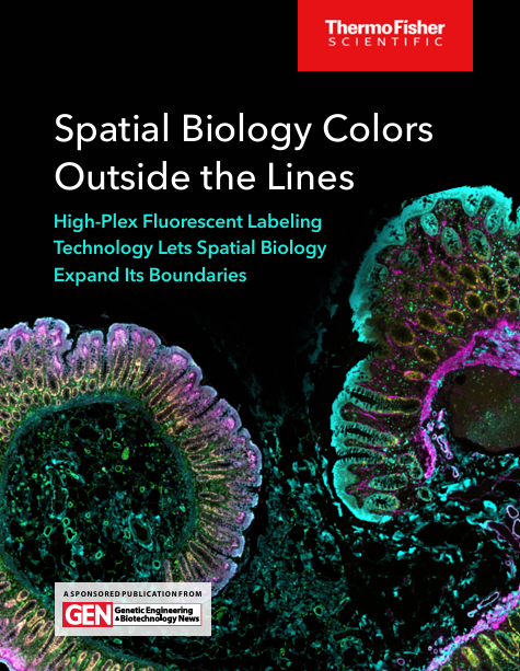
- Spatial Biology Colors Outside the Lines ー High-Plex Fluorescent Labeling Technology Lets Spatial Biology Expand Its Boundaries
会社カテゴリー:研究関連資材
主サービス提供地域:
製品・サービス詳細
Spatial Biology Colors Outside the Lines ー High-Plex Fluorescent Labeling Technology Lets Spatial Biology Expand Its Boundaries
サービスカテゴリー:分析、検査
You want to go spatial.
We’ll help you get there.
Breaking into spatial biology is easier than you may think
Spatial biology gives you the power to better understand how cells interact within tissue and how those interactions influence biological processes in healthy or diseased tissue.
Regardless of what platform you’re currently using, Thermo Fisher Scientific will collaborate with you to help get you started faster and more easily. Our offerings comprise high-quality, easy-to-choose, and easy-to-use reagents and antibodies, including Invitrogen™ Alexa Fluor dyes, all designed to help you save time and complete complicated experiments confidently.
Break into spatial biology today
A new spatial biology is in sight—literally. It is a more vivid, more detailed, and ultimately more informative spatial biology. It can distinguish between cell types that were once indistinguishable, and it can do so while capturing their spatial context. It can even pinpoint the subcellular locations of individual molecules. And it can accomplish these tasks with unprecedented precision because technology is now available that opens a new dimension beyond the usual three spatial dimensions. This new dimension may be called the “plex” dimension.
Plex refers to the number of fluorescence markers that are used with microscopy and other cell analysis platforms. Conventional platforms may accommodate just a handful of markers, constraining investigations of complex biological phenomena. But such investigations may require many markers.
Unfortunately, using more and more markers—and thereby shifting from low-plex to high-plex spatial biology—has been too difficult for most laboratories. They’ve balked at the need for special expertise, complicated workflows, and instrument upgrades. Fortunately, these difficulties can be overcome with new multiplex imaging technologies. For example, there are antibody panels that are compatible with streamlined workflows and automated imaging systems.
To learn more about these technologies, consult the articles in this eBook—especially the article describing organ mapping antibody panels. Also, be sure to read the articles that describe the kinds of spatial biology applications that are bound to become more common as high-plex technology becomes more accessible. Indeed, this technology is democratizing spatial biology.
GEN Interview with Oggie Golub, PhD,
Staff Scientist, Cell Biology R&D at Thermo Fisher Scientific
 OGGIE GOLUB is a cell biologist and microscopist with a doctorate degree in cellular and molecular biology from the University of Oregon. He has over eight years of R&D experience in advanced microscopy methods, including high resolution and high plex imaging using spectral detection. He leads a technical team working on the development of next-generation cellular analysis tools and workflows, with a focus on labeling and detection solutions for FFPE & cryopreserved tissue samples in spatial biology. |
GEN: What first piqued your interest in spatial biology and when did that happen?
Golub: At Thermo Fisher Scientific, we’ve been collectively working in earnest on spatial biology. But spatial is something in which I’ve personally always been interested. During my dissertation, I studied how asymmetric cell division gives rise to the cellular diversity that’s found in fully developed tissues, and that’s basically the fundamental theme of spatial biology—the complexity in cellular diversity of intact tissues.
On the technical front, fluorescence microscopy is a commonly used detection modality in spatial biology because it naturally lends itself to revealing the spatial organization of complex tissues. It was really the culmination of those two personal passions that really piqued my interest
in the field.
GEN: Building on that, what do you focus on, in terms of spatial, in your role at Thermo Fisher Scientific?
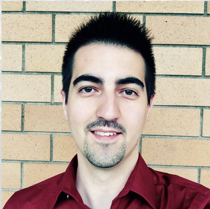
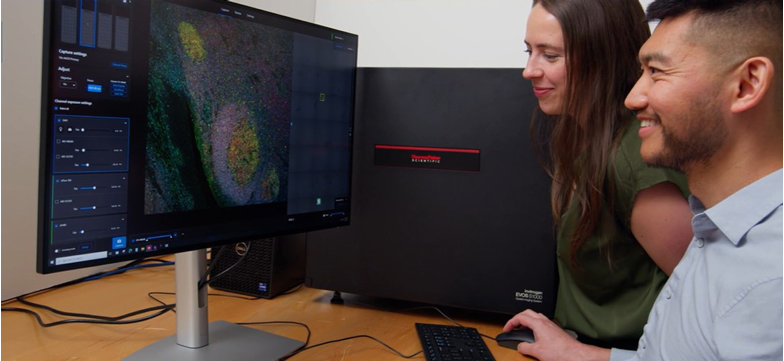
Golub: I am a staff scientist and R&D lead for spatial biology labeling and detection technologies. The focus of our entire group is to enable broader access to spatial biology through simplified workflows all the way from a tissue on a slide to an image that comes off an instrument. This includes sample preparation and labeling reagents, such as directly conjugated antibodies. We also have branched DNA technology with our ViewRNA kits that supports mRNA labeling, as well as our newest multiplex spectral detection platform—the EVOS S1000 spatial imaging platform.
EVOS S1000 spatial imaging platform
GEN: What’s the state of spatial biology today in your view?
Golub: It’s an interesting question because spatial biology is still rather new and rapidly evolving. There’s a lot of focus on plex and detecting the highest number of markers or targets possible. But when we speak to scientists who are using these technologies, they describe how the data itself is often quite overwhelming and it’s quite a struggle to do anything useful with that many markers.
The increase in plex also increases the complexity, scanning throughput, time to data, and overall cost of these experiments both on the reagent and instrument fronts. Therefore, we focused most of our efforts on specific areas to improve the workflows associated with high-plex fluorescence labeling and detection.
We also want to address existing pain points to eliminate barriers for new users, most of whom may not have the budgets or expertise to do these experiments today. Due to these barriers, spatial has still largely remained exclusive to relatively well-funded labs and core facilities with dedicated staff, extensive expertise, sample prep, and data collection and analysis capabilities.
Our aim really is to democratize this technology by taking away as many of these complexities as possible to make the technology accessible for all. It’s something that we internally refer to as the EVOS ethos. We try to simplify and remove complexity across the entire workflow, but specifically in labeling and detection.
GEN: What areas of biology do you think will most benefit from spatial in the next five years?
Golub: Immuno-oncology is the obvious answer because that’s where spatial is the primary application today. I think that will continue to increase, which should come as no surprise given that spatial provides invaluable insights into how cancerous tissues are organized, as well as how the immune system
responds to different treatments and disease progression. I also see a lot of potential in basic science applications, for example, in developmental biology to answer questions about transcriptomic and proteomic expression profiles during specific phases of development.
Spatial has a lot of potential to answer the question of where these markers are being expressed and at which times in development. As a result, I do see spatial biology being widely adopted and accessible for mid-plex protein or mRNA transcript on sectioned tissues. Cutting edge users will try to advance more towards 3D models and delve deeper into areas like metabolomics and maybe even epigenomics.
GEN: How might spatial change the course of cancer research in future?
Golub: Looking at the 2024 AACR conference topics, there was a strong focus on spatial technologies, and that’s presumably driven by the potential of spatial to inform molecular mechanisms of disease progression in cancer at the tissue level. But informing treatment potential is a much more challenging task because these assays will need to become a lot more standardized and more robust to be clinically validated. That’s where the big challenge lies but there are also some issues on the detection side with instruments needing to comply with specific types of regulatory standards.
The potential is certainly there for these technologies to make it into the clinic and inform treatments. I believe that low to mid plex fluorescence-based spatial platforms will soon supplement traditional histology techniques such as H&E staining but is very unlikely that they will supplant them. It will be another tool in the toolkit that could be used to inform personalized medicine at the molecular and cellular level.
GEN: What are some other examples of spatial research that excite you?
Golub: Spatial biology lends itself to the study of many biological processes because it’s really the study of the biological organization of intact tissues. I am excited for spatial to go beyond cellular interactions to elucidating potential microbe interactions as they pertain to both development and disease. We’ve learned a lot over the past decade, about the importance of the microbiome but it seems we still forget how dependent our biology is on microbial signaling cascades that inform both disease and development. That’s another potentially exciting area where spatial is likely to break into in the coming years.
GEN: What do you think users prioritize in a spatial platform?
Golub: Even though I’m a cell and molecular biologist by training, I’m really a technologist and microscopist at heart. I’ve spent a lot of time thinking about the technical aspects of these different platforms and which ones matter. Plex is certainly an important component of spatial and is generally the first spec that gets mentioned because traditional microscopy platforms that are capable of three to four plex are simply not sufficient in this regard. You need more markers to elucidate the variety of cells that are present in the tissue. And there are certain applications that truly do require a high number of markers—
The EVOS family of products empowers researchers, allowing them to focus on gathering data instead of instrument upkeep.
15, 20, maybe even 40 and beyond, but we have found that many researchers in spatial require 8–12 markers, which is the range supported by our new EVOS S1000 Spatial Imaging system. However, with increasing plex comes increased complexity and significant time investment to optimize the labeling conditions. To lower that barrier, we’re expanding our menu of primary antibody conjugates that are validated for use on FFPE preserved tissues so that users can have more ready-to-use validated antibodies that are basically available off the shelf.
We’ve also developed a new line of dyes, the Aluora Tyramide reagents, that are specifically formulated for multiplex immunofluorescence on FFPE, because we understand that validated reagents for labeling are immensely important, and some users don’t really have the desire or expertise or budgets to make their own. On the detection front, what we’ve heard is a clear need to simplify the workflow and make the imaging platforms faster and more approachable.
Existing solutions are either too complex or too expensive, or in some cases both, with some platforms requiring single-use consumables each time the user runs an assay. We’ve also heard a need for speed and high-resolution images because the downstream analysis depends heavily on high quality image data to perform accurate cell segmentation and feature extraction.
The newest member of the EVOS family, the S1000, uses high resolution optics and is built on top of a fast and flexible platform with an immensely intuitive user interface. Our hope is that the intuitive user interface will eliminate the need for extensive training and post-installation support and enable this technology to be basically accessible to every lab.
Our goal is to empower scientists with our products so they can spend more time collecting valuable data rather than maintaining and calibrating their instrument.
GEN: What are the current challenges in the field and how do you believe the field will get around them?
Golub: We’ve talked about the labeling challenges and complexity but there’s a big data problem. Since spatial biology deals with large areas of tissues across many, many markers, the size of the image datasets can easily approach tens and hundreds of gigabytes per experiment. And by the time you get done processing an entire study, you can easily end up with terabytes of data. It’s a logistical challenge for a lot of departments, both in terms of storage and organization, in addition to the computational power needed to work with those images.
The other piece is that the term “data” takes on multiple forms. Image data isn’t really quantitative by design so extracting high quality quantitative features from images remains immensely challenging. This too is partially limited by reagent availability because for segmentation, there are no dedicated pan-membrane markers to accurately elucidate cellular boundaries.
A lot of segmentation algorithms rely on nuclear counter stains, and these algorithms are only as capable as the information they have available—the stains and labels on the tissue. Artificial intelligence is a commonly prescribed panacea provided to the model, but AI models require training, and that training is only as good as either the user in supervised training or the labels that are available to it in unsupervised training.
How will the field get around these challenges? A part of the solution honestly lies in our hands. At Thermo Fisher Scientific we’re aware that the problem needs to be addressed and with any new technology, a lot of investigation and planning goes into that process.
For the data question, this is where advances in data science and applications of AI are likely to be the most needed. Addressing this problem requires really close and effective collaboration between biologists and data scientists in order to arrive at approaches that can enable efficient extraction of
biologically meaningful data from immensely complex spreadsheets of raw measurements.
|
|
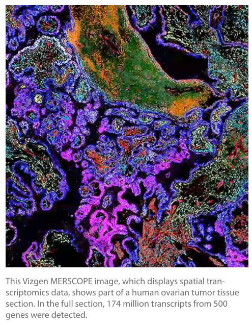
Spatial Biology: How It’s Transforming Single-Cell Genomics
A GEN-led panel conversation on spatial biology technology highlights the importance of community—not just among cells, but also among scientists
Spatial biology has all the hallmarks of the next omics revolution. For example, a key spatial biology technology, spatial transcrip
tomics, was named Method of the Year by Nature Methods in 2021. More generally, spatial biology is moving in a multiomics direction.
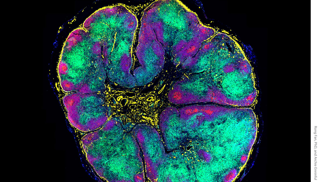
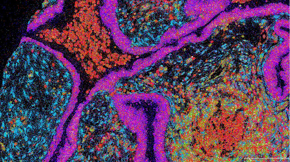
Shown above is a 52-plex immunofluorescence image of a whole human lymph node. Selected markers: DAPI (blue), CD20 (red), CD3e
(green), collagen IV (yellow). These markers identify nuclei, B cells, T cells, and vascular cells, respectively.
BY JULIANNA LEMIEUX, PHD
When the sober-minded describe spatial biology, they use words such as fledgling, growing, and maybe even exciting. But perhaps more enthusiasm—and words such as revolutionary and transformative—are in order. Yes, the basic idea behind spatial biology has been around for decades. Put simply, this is the idea that single-cell analysis should reflect how cells are organized in tissues. As familiar as this idea may seem, the toolkit to explore tissues spatially has been limited. But new technologies are making spatial biology a reality, ushering the field into a whole new era. Doesn’t that justify words that might otherwise seem hyperbolic?
 To convey the passion surrounding spatial biology, GEN hosted a panel discussion titled, “How Spatial Biology is Revolutionizing Single Cell Genomics.” It originally aired on October 18 as an episode of GEN Live, our science talk show. And it is still available to anyone who wants to hear about the current status of spatial biology, the impact the technology is making on research (and perhaps one day, patients), and the challenges it faces as it moves forward.
GEN revisits the panel discussion here. Moreover, in sympathy with the basic idea behind spatial biology—that is, the idea of contextualization—we provide some additional background and commentary. We include direct quotes from the two panelists, namely, Rong Fan, PhD, professor of biomedical engineering at Yale University, and Nicholas Banovich, PhD, associate professor in the Integrated Cancer Genomics Division at the Translational Genomics Research Institute (TGen). Our aim is to highlight ongoing trends, challenges that are being overcome, and future possibilities that inspire people to drive spatial biology forward.
Then and now
 The discussion kicked off with a broad view of spatial biology’s evolution. To understand how the technology arrived at where it is today, Fan started with a historical perspective, going back to the work of Robert Hooke (in the 1600s) and his early descriptions of how cells appeared when viewed through the microscope. Then, Fan moved to more modern localization techniques used to investigate proteins and nucleic acids spatially in tissues, including immunostaining and fluorescence in situ hybridization. Now, with the newest wave of spatial technologies, researchers have the ability to dig into mechanisms and function.
Fan noted that analyzing gene expression is currently the centerpiece of spatial. One reason for that, Banovich noted, is the digitization of the signal. The analysis of data in a way that uses the existing genomic tool set seems to be what allowed spatial biology—particularly spatial transcriptomics—to become broadly useful.
“Whether you’re talking about imaging-based platforms or sequencing-based platforms, being able to churn these through the genomics infrastructure has really led to this broad adoption of the technologies,” Banovich remarked. And because the transcriptomics signal is straightforward to digitize—compared to proteomics or epigenomics signals—it was the first to emerge as the dominant technology.
Fan added that there will be more access to investigate above (proteins and metabolites) and below (the genome and epigenome) the RNA in the cell. And that will generate a new question: Can we link all of them together?
Where are we now?
“This feels to me very much like the single-cell world of 2016 or 2017,” Banovich said, “where everybody is ‘out in the woods,’ trying to figure out exactly what they can eke out of these technologies.” He explained that there are a lot of researchers wrapping their heads around what they can do with these data. Getting relational information, Banovich continued, is the gold mine of spatial data. But there is a lot of work going into understanding how to use the relational information, figuring out what tools are available to answer those questions, and designing the computational tools that still need to be built.
In the early days of single-cell biology, the sharing of methods and experiences was commonplace. And Banovich expressed his hope that this kind of sharing would be the norm in spatial biology, too. One major difference, he observed, is that single-cell biology was heavily dominated by a single technology. As a result, single-cell biology was given to uniformity. In contrast, there is a huge diversity of spatial technologies, even with respect to a single modality such as transcriptomics.
What they are constantly thinking about in Fan’s laboratory is not just how to standardize, but how to integrate data from different modalities. And even when the data from different modalities is there, he said, there will still be a lot of work in understanding how to put them together. But people are moving in this direction now. He noted that computational biologists are playing a key role in this aspect of spatial.
Collaboration is key
Given the diversity in technologies, there is an increased need among researchers to share openly, making community driven initiatives incredibly valuable. One community that has
developed over the past few months is the Global Alliance for Spatial Technologies (GESTALT). It was started by Ioannis Vlachos, PhD, co-director of the Bioinformatics Program, Cancer Research Institute, Beth Israel Deaconess Medical Center; Luciano Martelloto, PhD, associate professor at the University of Adelaide; and Jasmine Plummer, PhD, associate member, St. Jude Faculty and director, Center for Spatial OMICs.
GESTALT holds meetings online that allow spatial users from around the world to come together, share knowledge and challenges, and collaborate. Over 100 people attended the first online meeting. The founders hope that it will be a catalyst in advancing spatial technologies. The focus is on collaboration, knowledge sharing, and innovation that will serve to amplify the impact of spatial technologies across sectors.
GESTALT is a space to foster productive dialog for everyone, provide valuable feedback for active users, advocate for best practices for presenting data, and pool growing expertise to address chal lenges and foster innovations. The group plans to establish working groups to push the needle on specific topics such as standards and best practices, assay optimization, and data analysis.
The spirit of open science is a terrific part of the single-cell community, Fan stressed. And because the single-cell community comprises many of the researchers moving to spatial, that collaborative spirit will be brought into the spatial world as well.
The Banovitch lab at TGen specializes in multiomics, with a particular focus on diseases, and identifying genetic variation associated
with changes in gene regulatory phenotypes. One area of research is uncovering the molecular mechanisms underlying pulmonary
fibrosis. Here, spatial transcriptomics examines fibrotic lung disease using the 10X Genomics Xenium platform. Each point is a
transcript, and they are colored by the cell type typically associated with that gene.
Bringing it all together
One thing that the spatial community would benefit from is a reference. Fan suggested that the spatial community might follow the example set by the human genome project. Once a reference genome was built, researchers could map everything to the reference genome. In the spatial community, an analogous effort would help
researchers make sense of observations and data.
Banovich indicated that he appreciates the efforts of the atlas community over the past couple of years. These efforts include the building and unification of cell atlases across different organs. Once a certain point is reached, researchers could map spatial data to single-cell reference data, unifying datasets and minimizing batch effects. But that, he admitted, poses a huge challenge.
Moving from the collection of spatial data to the generation of insights and knowledge is a long way to go, Fan said. And practical challenges arise from spatial biology’s multidisciplinary nature. To illustrate this point, Fan offered the following example: A pathologist who has decades of experience in tissue embedding knows how to process tissue specimens but is unlikely to know the omics side of the technology. This intrinsic challenge requires people to work together. If people want to succeed, Fan maintained, they need people with extremely diverse backgrounds working together to overcome those barriers.
Magic wand
If given a magic wand, what would Fan and Banovich wish for to move spatial forward? An increase in plex, sensitivity, or resolution? Multiomics in one run? Or more sophisticated bioinformatics?
Banovich answered, laughing, that if the wand is really magic, he would change them all! The reason it is so hard to pick one, he explained, is because it is too early to know which of these things will really elevate spatial to the next level. He also contributed this brief self-dialogue: “Would I love an increase in plex? Absolutely. Does that come with a hit to sensitivity? Probably.”
He said that he would be incredibly excited about a 10,000-plex spatial transcriptomics maps. But he added that if such maps were to come with a 75% sensitivity drop, he would immediately feel less excited. A 10,000-plex capability today, with no drop in sensitivity, would change the game for the spatial platforms.
Fan told GEN that he “wants everything, for sure.” And that is not too far out of range; it is becoming possible. He emphasized that the field should embrace artificial intelligence and machine learning for computational data. The real revolution of the field, he asserted, will come when computer scientists, and artificial intelligence researchers, join the force to write the next chapter.
|
How to pick antibodies for spatial biology
|
|
 Presenter:
さらに詳しい情報は、ページ右上の青い「ダウンロードはこちら>」ボタンをクリックし、ダウンロードしてください。
Spatial Biology Colors Outside the Lines