ライフテクノロジーズジャパン株式会社 バイオサイエンス事業部
- HOME
- ライフテクノロジーズジャパン株式会社 バイオサイエンス事業部
- 製品・サービス詳細
- Spatial Imaging Demystified Latest Tools, Techniques, and Workflows Shaping the Future of Spatial Biology
会社カテゴリー:研究関連資材
主サービス提供地域:
製品・サービス詳細
Spatial Imaging Demystified Latest Tools, Techniques, and Workflows Shaping the Future of Spatial Biology
サービスカテゴリー:分析、検査
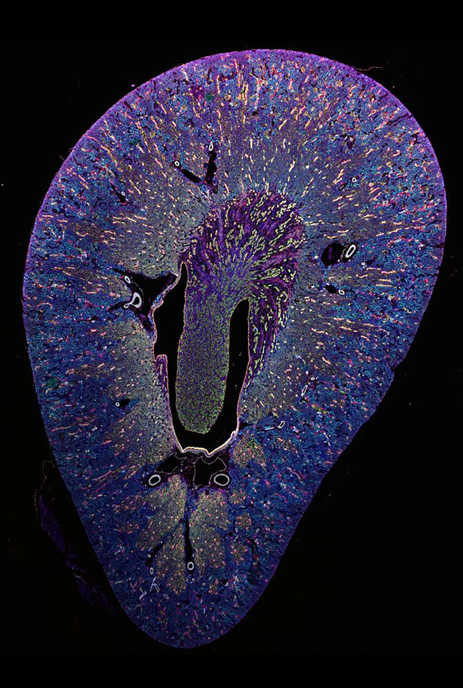 Introduction
The way we study biology is changing. Advances in digital imaging, multiplexed detection, and high-resolution visualization are transforming our understanding of biology, and producing more insights into biological processes and mechanisms of disease.
This eBook offers a comprehensive look at the tools and techniques involved in spatial imaging and explores topics including:
• Foundational concepts of spatial biology
• Comparison of imaging techniques
• Emerging applications across research domains
• Experimental design considerations
• Advances in multiplex imaging solutions
Understanding Spatial Biology
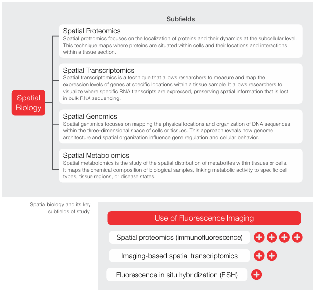 Spatial biology is the comprehensive study of biomolecules and cells in their native context. By combining imaging, molecular profiling, and computational analysis, it reveals insights into tissue structure, cell-cell interactions, neighborhoods, and microenvironments within their natural tissue environments. Spatial biology comprises subfields such as spatial transcriptomics, spatial proteomics, spatial genomics and spatial metabolomics.
Spatial Imaging:
The Key to Visualizing Biology in Context
Spatial imaging is a set of techniques used to understand spatial biology. They combine molecular analysis with high-resolution imaging to map cells, proteins, and gene expression directly within intact tissue. Spatial imaging preserves tissue architecture, making it possible to visualize biology in its native context
Immunofluorescence imaging is a widely used spatial biology technique, enabling visualization of target molecules within cells and tissues.
Fundamentals of Spatial Imaging
In biology, context is essential to understanding complex biological systems. Spatial imaging captures crucial dimensions—revealing how cells are organized, how tissues are structured, and where key proteins and signals reside within their native environment. By preserving tissue architecture while capturing highresolution molecular and cellular data, it enables researchers to visualize where and how different components interact. Whether it’s mapping immune cell infiltration in tumors, exploring the tissue-specific traits between drug responder and non-responders, analyzing brain tissue organization, or studying developmental biology, spatial imaging reveals patterns and relationships that were not possible to understand until now.
Spatial imaging refers to technologies that capture molecular and cellular information within the intact architecture of tissue. Unlike methods that analyze dissociated cells or homogenized samples, spatial imaging maintains the physical location of cells, proteins, and other markers—allowing insight into not just what’s present, but where it’s located and how it interacts with its surroundings.
At its core, spatial imaging integrates two key dimensions:
• Molecular specificity - Detection of proteins, nucleic acids, or other biomarkers using techniques like immunofluorescence, in situ hybridization, or antibody panels.
• Spatial resolution - Visualization of these markers within the structural context of the tissue, from cellular detail to tissue-wide patterns.
Core concepts include:
• Tissue architecture - The structural organization of cells and extracellular matrix within a biological sample.
• Co-localization - Identifying multiple markers in the same region or cell to study functional relationships.
• Multiplexing - Imaging multiple biomarkers simultaneously or sequentially in a single sample.
• Segmentation and quantification - Using image analysis to define cells, regions, and quantify marker expression.
As research questions grow more sophisticated, especially in oncology, neuroscience, and immunology—spatial imaging is no longer optional. It’s becoming a core tool for generating insights that are biologically critical, more meaningful and more translationally relevant. Together, these components allow researchers to ask complex biological questions about cell function, communication, and spatial heterogeneity—questions that static or bulk methods can’t address.
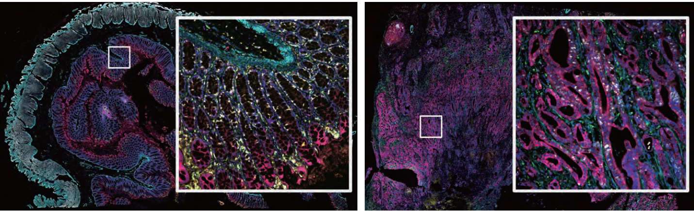
Figure 1. Detecting more targets helps provide more details about the tissue microenvironment and highlights the
complexity of biological systems within tissues. Images of normal colon (left) and adenocarcinoma of colon tissues (right)
stained with the 9-plex colon panel on the EVOS S1000 Spatial Imaging System. Multiplex immunofluorescence staining enables
information to be collected about the localization and interaction of biomolecules and cells within the tissue microenvironment.
Spatial Imaging Techniques
The two most widely used methods for multiplex fluorescence-based spatial imaging are spectral imaging and cyclic imaging.
Spectral Imaging: suitable for mid-plex experiments and translational research
• How it works: Captures the full emission spectrum from each fluorophore and uses software to unmix overlapping signals.
• Why it matters: You can use multiple fluorophores at once—even ones with very close emission wavelengths—because spectral unmixing separates them computationally.
• Advantages:
– Faster - Everything is stained and imaged in one shot.
– Gentler - Preserves tissue integrity and less photobleaching.
– Flexible - While typically used for one-shot imaging, spectral imaging also supports cyclic labeling when higher plex levels are needed, eliminating the dependency of just labeling in cycles
• Limitations:
– Multiplexing is typically capped at ~6–9 markers.
– Limited by how well the system can resolve overlapping spectra.
More complex assay design
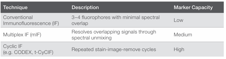
Cyclic Imaging: suitable for high-plex experiments and discovery research
• How it works: Stain a few markers → image → chemically or physically strip the fluorophores → repeat for new markers and image the markers in repeated cycles of 2-4 fluorophores per cycle
• Why it matters: You’re not limited by spectral overlap, because you’re only imaging a few markers at a time.
• Advantages:
– Much higher multiplexing (20+ markers is common).
– Simpler panel design.
• Limitations:
– Time-consuming due to multiple rounds of staining and imaging.
– Tissue degradation and registration errors can accumulate over cycles.
– More demanding sample prep and instrument handling.
– Data handling may be challenging due to computing power.
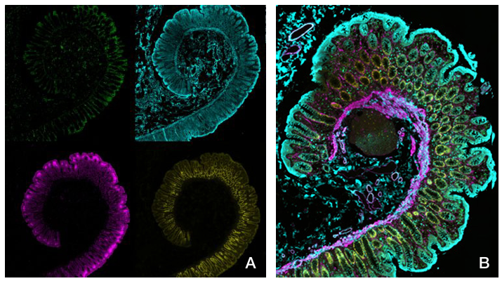
Fig. 2 More colors show more details about the tissue microenvironment.
(A) Staining of healthy colon tissue with four different labels. A limited number of targets results in limited information available for understanding the tissue microenvironment.
(B) Staining of healthy colon tissue with nine different labels spectrally unmixed. Multiplex immunofluorescence staining enables information to be obtained about the localization and interaction of biomolecules and cells within the context of a tissue.
Spatial Biology and the Shift Towards Contextual Data for Disease Research
|
Across biology and medicine, there’s a growing recognition that spatial context isn’t just helpful, it’s essential. Nowhere is this shift more evident than immuno-oncology, neuroscience, and pathology. In these fields, understanding cell locations and how they interact within the tissue microenvironment can make or break an insight and impact progress towards disease understanding and development of new therapies. In immuno-oncology, spatial biology is unlocking the complexity of the tumor microenvironment (2,3). It reveals whether immune cells are infiltrating or excluded, how close they are to tumor cells, and what checkpoint markers they express—all of which impact response to therapy. Spatial biology addresses questions such as: • Are cytotoxic T cells in contact with tumor cells or excluded by stroma/myeloid cells? • What location are PD-L1, LAG-3, TIM-3 expressed—at interfaces, inside nests, or in perivascular niches? • Do tertiary lymphoid structures (TLS) form, and which B cells/T cells subsets populate them? Spatial imaging turns the tumor microenvironment into measurable features such as interfaces, niches, and gradients. This spatial context in turn enables researchers and clinicians to move beyond cell counts and into actionable tissue architecture and higherresolution investigations. In neuroscience, the importance of spatial relationships is foundational. Brain function depends on precise structural organization and features such as layers, Pathology is inherently spatial. Digital and spatial tools are transforming static tissue evaluation such as IHC into data-rich, multi-parametric analysis. Spatial imaging allows pathologists to layer molecular insights on top of histological structures, enabling better comprehension of molecular disease drivers to ultimately guide diagnostics and better therapeutics for the right patients. This next level integration is allowing improvements including: • Precise resolution of overlapping morphologies • Standardized quantification of attributes such as macrophage states and stromal features • Region-aware profiling for morphology-guided biomarker quantitation Spatial imaging complements diagnostic workflows with scalable, quantitative context, accelerating translational studies and paving the way for validated clinicalgrade assays. |
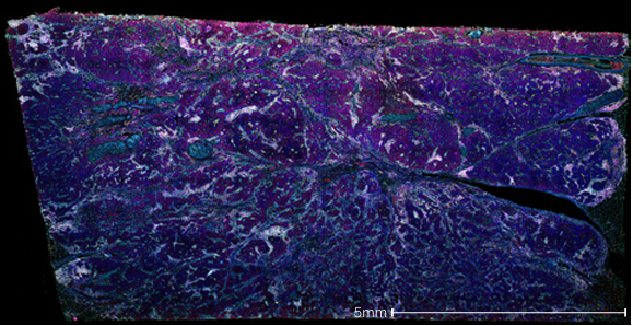
Figure 3. Human invasive ductal carcinoma of breast tissue. Image was processed and stained with the Aluora spatial amplification 8-plex assay using Invitrogen’s EVOS S1000 Spatial Imaging System.
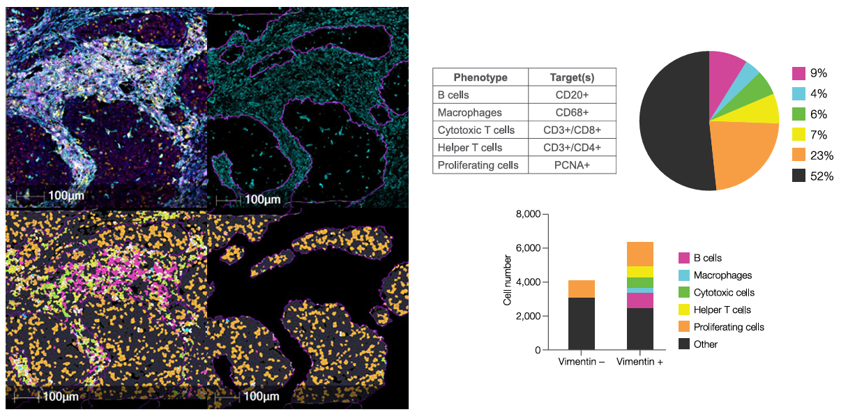
Figure 4. Single cell segmentation and phenotyping revealing spatial distribution of immune cell subpopulations. Summary Spatial imaging has expanded classic fluorescence into a multiplex, quantitative, and context-aware discipline. By preserving where biology happens—and measuring many targets at once—it delivers the maps researchers and clinicians need to understand interactions, compare cohorts, and make decisions. In immuno-oncology, neuroscience, and pathology alike, an imaging-first approach connects molecular state to tissue architecture in ways that bulk assays cannot.
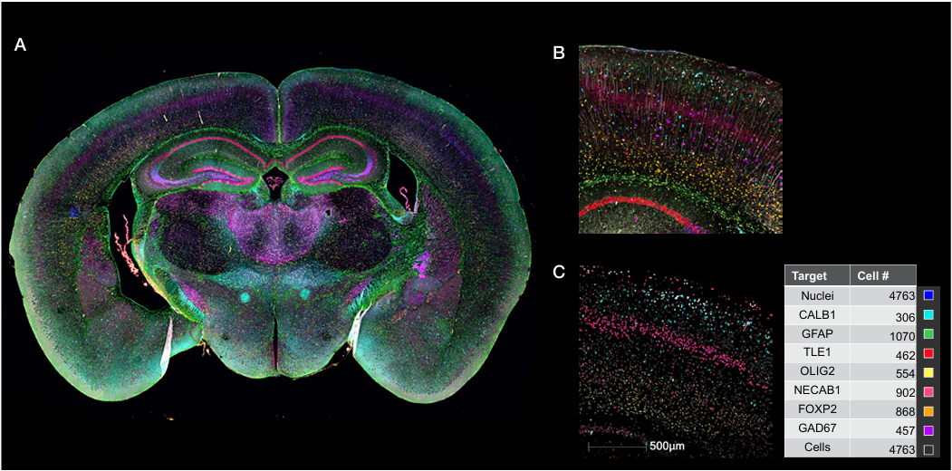
Figure 5. Mouse coronal brain tissue section. A) Composite image, B) Zoomed-in inset, C) Quantification of target positive cells. Tissue was stained with Aluora spatial amplification 8-plex assay and imaged using Themo Fisher’s EVOS S1000 Spatial Imaging System coupled with HALO ® analysis software. |
Designing Robust Spatial Imaging Experiments
While high-plex labeling with fluorescent dyes requires careful, multi-step workflows—including staining, stripping, and imaging—these processes enable the generation of richly detailed spatial data. Advances in imaging platforms are continually improving usability and throughput, helping researchers overcome earlier challenges in spatial proteomics. As with any sophisticated technique, attention to methodological precision ensures consistent, high-quality results that deepen biological insight. These challenges can include:
1. Specimen/sample preanalytical variation
2. Panel design and antibody/probe validation
3. Image acquisition
4. Segmentation and cell typing
5. Reproducibility and harmonization
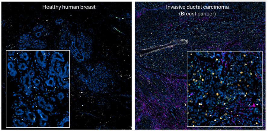
Spatial imaging offers a holistic view of complex biological systems
Spatial imaging turns tissue sections into quantitative maps—but only if the experiment is designed to withstand real-world variability and analysis complexity.
1. Preanalytical variation can be minimized by addressing source variability, fixation parameters, sectioning strategy, and autofluorescence.
2. Multiplex panel validation can be achieved by verifying antibody sensitivity and selectivity, optimizing the order of method cycles, testing for reagent cross-reactivity and carryover, and using controls at all stages.
3. Image acquisition can be improved by optimizing slide preparation, exposure and focusing, illumination, and tracking all metadata to quantify and control variations and set acceptance criteria.
4. Segmentation and cell typing can be enhanced using whole-slide visualization, combining marker intensity, co-expression, and morphology, and assembling acceptance or exclusion criteria.
5. Batch reproducibility and harmonization can be achieved by including cross-patient sample groups, using reference materials, correcting or removing autofluorescence, and cross-instrument testing.
Designing a successful spatial imaging experiment requires thoughtful planning and rigorous validation at every stage—from sample preparation to data harmonization.
While the complexity can be daunting, implementing optimized workflows, validated reagents, and well-designed panels can dramatically reduce variability and improve reproducibility. For many researchers, striking the right balance between biological insight and operational efficiency begins with carefully selecting how many markers to image and which platform best supports those goals.
Mid-Plex Imaging: Making Multiplexing More Accessible
In spatial imaging, more isn’t always better. While high-plex platforms promise dozens of markers, they often require complex workflows, long acquisition times, and extensive data processing. At the other end of the spectrum, traditional low-plex imaging can miss the biological context researchers need.
This is where mid-plex imaging offers an elegant solution. By imaging five to nine markers in a single scan, mid-plex imaging delivers highly multiplexed insights while keeping workflows streamlined, reproducible, and cost-effective. It’s fast enough for routine studies, powerful enough for deep biological exploration, and flexible enough to adapt to evolving research questions.
The EVOS™ S1000 Spatial Imaging System was developed with this shift in mind. By pairing spectrally optimized dyes, validated antibodies and reagents, and intuitive software, it empowers researchers to move from panel design to high quality spatial data without the trade-offs of high-plex cycling.
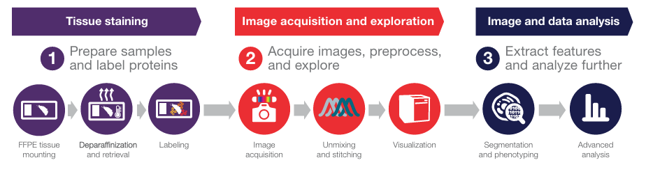
1) Spatial imaging workflow
The EVOS S1000 solution supports a complete path from FFPE tissue to quantitative single-cell readouts. Multiplex spectral imaging workflow overview:
1. Sample prep & labeling
2. Image acquisition and exploration
3. Segmentation, phenotyping & downstream spatial analysis
2) Instrumentation
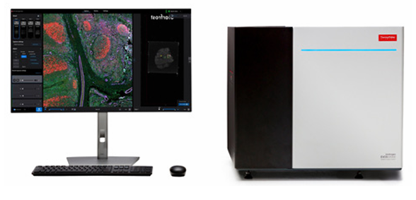
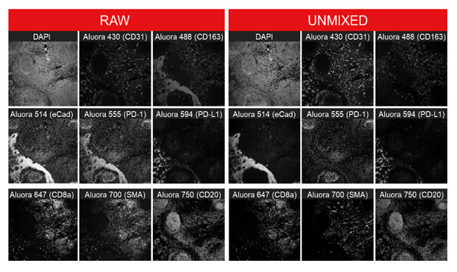
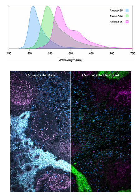
EVOS™ S1000 Spatial Imaging System is purpose built for multiplex immunofluorescence (mIF), acquiring up to nine channels simultaneously in a single round—preserving tissue while avoiding the dependency on iterative staining and imaging cycles. Integrated spectral unmixing is executed during acquisition, resolving overlapping emission spectra, and outputting quality metrics.
Key capabilities:
• Nine-color single-scan imaging at high resolution for protein localization and neighborhood mapping
• Compatible with various tissue labeling methods
• Preserves tissue integrity due to fewer imaging cycles.
• Automated, high quality spectral mixing
Watch EVOS S1000 spectral unmixing in action.
3) Spatial imaging reagents
Thermo Fisher Scientific provides three complementary labeling options plus mounting media, designed to pair with the EVOS S1000 and other spatial imaging systems.
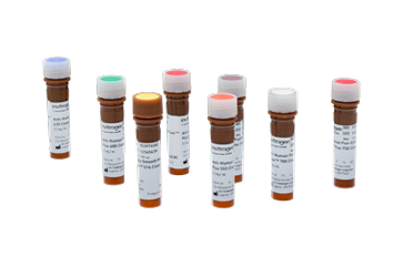 Alexa Fluor™ Conjugated primary antibodies.
Conjugated primaries remove the need for speciesspecific secondaries and speed up the staining workflow. Designed for one-step labeling of FFPE tissues, Invitrogen ready-to-use antibodies are conjugated to Alexa Fluor or Alexa Fluor plus dyes, ideal for tissue imaging, suitable for single or multiplex staining, and use with conventional and spectral imagers.
When to use: You need to detect numerous targets simultaneously using conjugated primary antibodies for multiplexing experiments. These antibodies have been tested on tissue samples and are available in formats compatible with the EVOS S1000 Spatial Imaging System and other microscopy platforms.
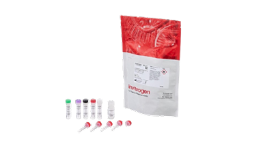 ReadyLabel™ Antibody Labeling Kits.
Do-it-yourself conjugation kits with popular Alexa Fluor/Alexa Fluor Plus dyes creates primary conjugates where off-the-shelf fluorescence options do not yet exist. No antibody purification required – they can be used even in the presence of carrier proteins. These labeling kits ensure high signal-to-background ratios.
When to use: You already have a validated primary, but no pre-conjugate exists; need rapid turn-around with minimal hands-on time and no need for prior conjugation experience.
 Aluora™ Spatial Amplification Kits.
Enzyme-mediated signal amplification with exceptionally bright fluorophores; the covalent fluorophore deposition is resistant to stripping, enabling reuse of same-species primaries across rounds without cross-reactivity. Available as individual dyes and HRP kits for anti-mouse, anti-rabbit, or streptavidin-conjugated primaries.
When to use: Low-abundance targets, limited primary supply, or challenging backgrounds. They have demonstrated lower exposure times and improved signal-to-noise ratios when compared to conventional secondary or other amplification chemistries in FFPE.
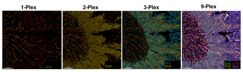
More colors, greater detail. Unmixed multifield region of an axial Murine kidney FFPE sample labeled with 8 Aluora dyes. Panels 1-4 show the same area of interest and illustrate that increasing the number of labeled targets within a sample provides greater detail. The images were captured using the 20x objective on the Invitrogen EVOS S1000 Spatial Imaging System.
Putting it together
|
A reference imaging workflow Practical EVOS S1000 run (9-plex): – Mount, deparaffinize, retrieve antigens. 1b. Label (these labeling methods can be mixed and matched): – Option A: Conjugated primaries - single-step 2. Controls & unmixing: The EVOS S1000 Spatial Imaging System software facilitates 3. Scan once: Acquire 9-channel composite; spectral unmixing runs during acquisition. 4. Export & analyze: Save unmixed, stitched OME-TIFF; segment cells; quantify phenotypes; |
|
|
Expected outcomes using these imaging tools and techniques • Higher-plex in fewer cycles: Nine fluorophores per scan; reduce handling, Take the EVOS™ S1000 Spatial Imaging & Ecosystem 3D Tour! The EVOS S1000 system and its validated antibodies and reagents offer a coherent, |
 |
Summary
As spatial imaging continues to evolve, researchers face an expanding spectrum of options, from low-plex techniques focused on simplicity to high-plex platforms capable of deep molecular profiling. Increasingly, however, many laboratories are finding that mid-plex imaging offers the ideal balance: enough markers to capture biological complexity, but with workflows that remain efficient, reproducible, and scalable.
The promise of spatial imaging lies not just in the technology itself but in what empowers researchers to achieve:
• Deeper understanding of cellular neighborhoods
• Better interpretation of tissue microenvironments
• New opportunities for biomarker discovery and translational breakthroughs
The landscape is moving fast. Overall, this pivot toward spatial biology reflects a broader
trend: moving from isolated signals to system-level understanding. This new contextual
data is not just enhancing existing workflows—it’s redefining the future of research and
clinical discovery.
References:
1. https://assets.thermofisher.com/TFS-Assets/BID/Application-Notes/evos-s1000-spatial-imaging-system-app-note.pdf
2. Galon J, Costes A, Sanchez-Cabo F et al. (2006) Type, density, and location of immune cells within human colorectal tumors predict clinical outcome. Science 313(5795):1960–1964. doi: 10.1126/science.1129139
3. Fridman WH, Pagès F, Sautès-Fridman C, Galon J (2012) The immune contexture in human tumours: impact on clinical outcome. Nat Rev Cancer 12:298–306. doi: 10.1038/nrc3245
Explore additional resources:
https://documents.thermofisher.com/TFS-Assets/BID/brochures/spatial-imaging workflow-selection-guide.pdf